Fiji Imagej Citation
Joubert syndrome-associated mutations in the human DUF3719 protein FAM149B1 as well as XBX-4 elongate cilia. If you need any help please first take a look at the.
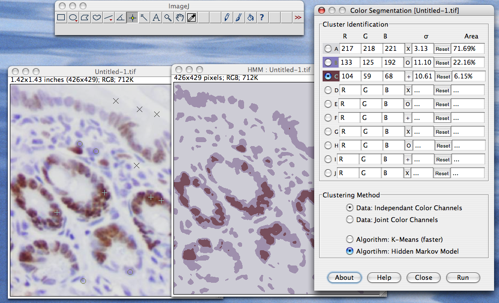
Color Segmentation
The Gene Ontology GO knowledgebase is the worlds.
Fiji imagej citation. Maurya and Sengupta show that the C. Citation needed The interactive visualization capabilities of 3D Slicer include the ability to display arbitrarily oriented image slices build surface models from image labels and hardware accelerated volume rendering. Diagnostics 2021 11 1948 2 of 10 However these studies focused on line scans or single measurement points through the macula which may not be sufficient to.
They work well on tiles and other smooth surfaces but do not stick well to rougher materials like brick or wood because they are unable to form an air-tight seal. The mission of the GO Consortium is to develop a comprehensive computational model of biological systems ranging from the molecular to the organism level across the multiplicity of species in the tree of life. The choroidal vasculature should be evaluated in three-dimensional 3D rather than from a two-dimensional scan.
DeconvolutionLab2 The remasterized Java deconvolution tool. Z-projected Max intensity was performed to convert the stacks into 2D images. The analyzed cells were randomly selected but we avoided choosing pavement cells adjacent to guard cells and hair cells because their shapes were distorted.
Suction cups are widely used to attach objects to surfaces in bathrooms and kitchens. Using ImageJFiji we manually traced the contours of 30 pavement cells and 30 subepidermal palisade cells per leaf. The blacklegged tick Ixodes scapularis a species of significant importance to human and animal health harbors an endosymbiont Rickettsia buchneri sensu stricto.
DeconvolutionLab2 is freely accessible and open-source for 3D deconvolution microscopy. With ImageJFiji these two readouts can be obtained using the Analyze Particles function which only works on binary 2D images. The images were quantified using ImageJ Fiji.
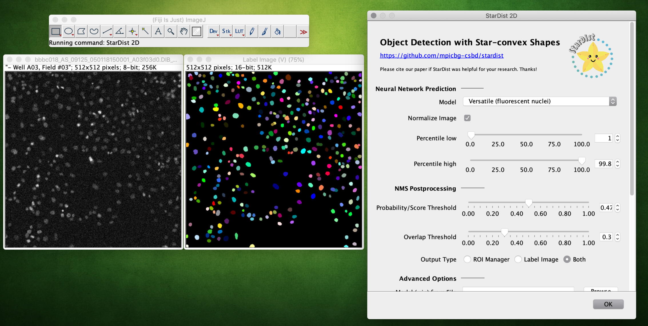
It can be linked to well-known imaging software platforms ImageJ Fiji ICY Matlab and it runs as a stand-alone application. Metabolic reprogramming in tumors is an adaptation that generates vulnerabilities that can be exploited for developing new therapies. This is the ImageJFiji plugin for StarDist a cellnuclei detection method for microscopy images with star-convex shape priorsThe plugin can be used to apply already trained models to new images.
A region without any cells were selected and used to subtract background fluorescence. This means that our image processing protocol should include both z-projection and binarization using a threshold these two treatments being compatible with our set up. IDL short for Interactive Data Language is a programming language used for data analysisIt is popular in particular areas of science such as astronomy atmospheric physics and medical imaging.
The backbone of our software architecture is a library that contains the number-crunching elements of the. All image processing steps discussed here and complementary suggestions for. DeepImageJ is a user-friendly solution that enables the generic use of pre-trained deep learning models for biomedical image analysis in ImageJ.
Both the whole construct and the fusion region was selected respectively using the drawing tool and the integrated density was measured using the built in ImageJ functions. GFP 470 22 nm excitation 510 42 nm emission and RFP 531 40 nm excitation 593 40 nm emission filter cubes were used to capture green or red fluorescence. Identify synergism between ABL allosteric inhibitors and lipophilic statins to impair metastatic lung cancer cell outgrowth and colonization leading to increased survival in mouse models of advanced disease.
ImagejFiji编写Python脚本Jython ImageJ weixin_49567369. For Sholl analysis a 20 objective NA 08 was used and Z-series stacks with a step size of 1 µm were recorded. The workflow is based on the open source software Fiji but its principles can be applied with other software packages.
The endosymbiont is cultivable in cell lines isolated from embryos of Ixodes ticks. Context-specific tuning of this kinase cascade may allow precise modulation of cilia architecture. The deepImageJ environment gives access to.
Citation needed IDL shares a common syntax with PV-Wave and originated from the same codebase though the languages have subsequently diverged in detail. Citation needed 3D Slicer also supports a rich set of annotation features fiducials and measurement widgets customized color maps. Citation needed Slicers.
The ImageJ wiki is a community-edited knowledge base on topics relating to ImageJ a public domain program for processing and analyzing scientific images and its ecosystem of derivatives and variants including ImageJ2 Fiji and others. We then examined the cell size and other shape-related indices using the Area Shape descriptors and. ImagejFiji编写Python脚本Jython ImageJ weixin_49567369.
The dendrites of neurons were traced with the semi-automated Simple Neurite Tracer plugin. Indeed as our tissue of interest is a monolayer z-projection should not affect. See the main repository for links to our publications and the full-featured Python package that can also be used to train new models.
The symbiont is largely restricted to the ovaries but all life stages can harbor various quantities or lack R. An EVOS onstage incubator was used for live cell experiments and images were quantitated using ImageJFiji 49 50 see supplemental methods supplemental appendix. From Cell Imaging to Data Analysis.
Diagnostics 2021 11 1948 2 of 9 vessels. Researchers have been searching for ways to improve these cups by studying how octopuses remora fish and other sea animals use muscle-powered. Cell migrationinvasion wound healing assay scratch assay transwell assay spreading assay live cell imaging data analysis ImageJFiji.
Here Luttman et al. Fluorescent intensity in the fusion region was quantified using ImageJ software version 61 Fiji National Institutes of Health. U-Net is a generic deep-learning solution for frequently occurring quantification tasks such as cell detection and shape measurements in biomedical image.
Pijuan J Barceló C Moreno DF Maiques O Sisó P Marti RM Macià A and Panosa A 2019 In vitro Cell Migration Invasion and Adhesion Assays. Elegans XBX-4 DUF3719 protein acts upstream of the CCRKRCK cascade to regulate sensory cilia structure.
Imagej Fiji Omero Microscopist Co Uk

Checker Checker Amap Dev

![]()
Citing
Fiji An Open Source Platform For Biological Image Analysis Nature Methods

Measurements In Imagej Fiji A Measurements Of Cell Width Line A Download Scientific Diagram

Development Of Labelstorois Plugin To Analyze Label Images In Download Scientific Diagram
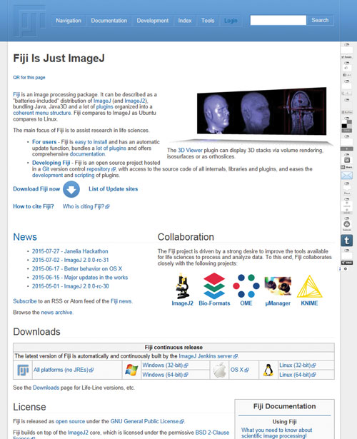
Fiji Is Just Imagej Best Of The Web

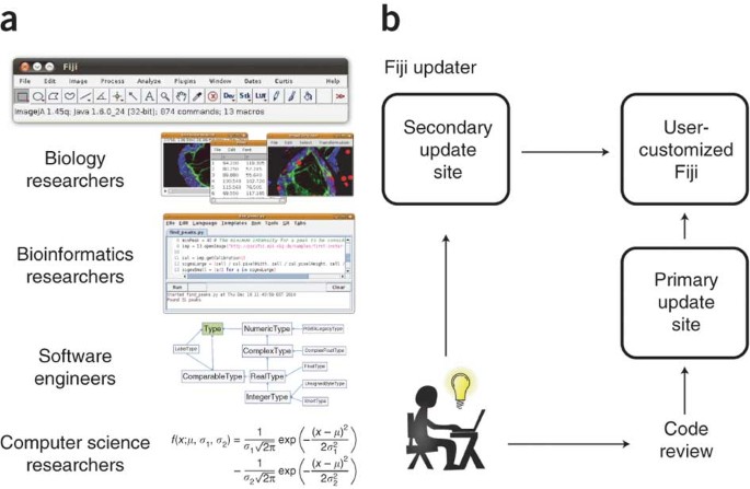
Fiji Imagej With Batteries Included
Stardist

Fiji Imagej With Batteries Included
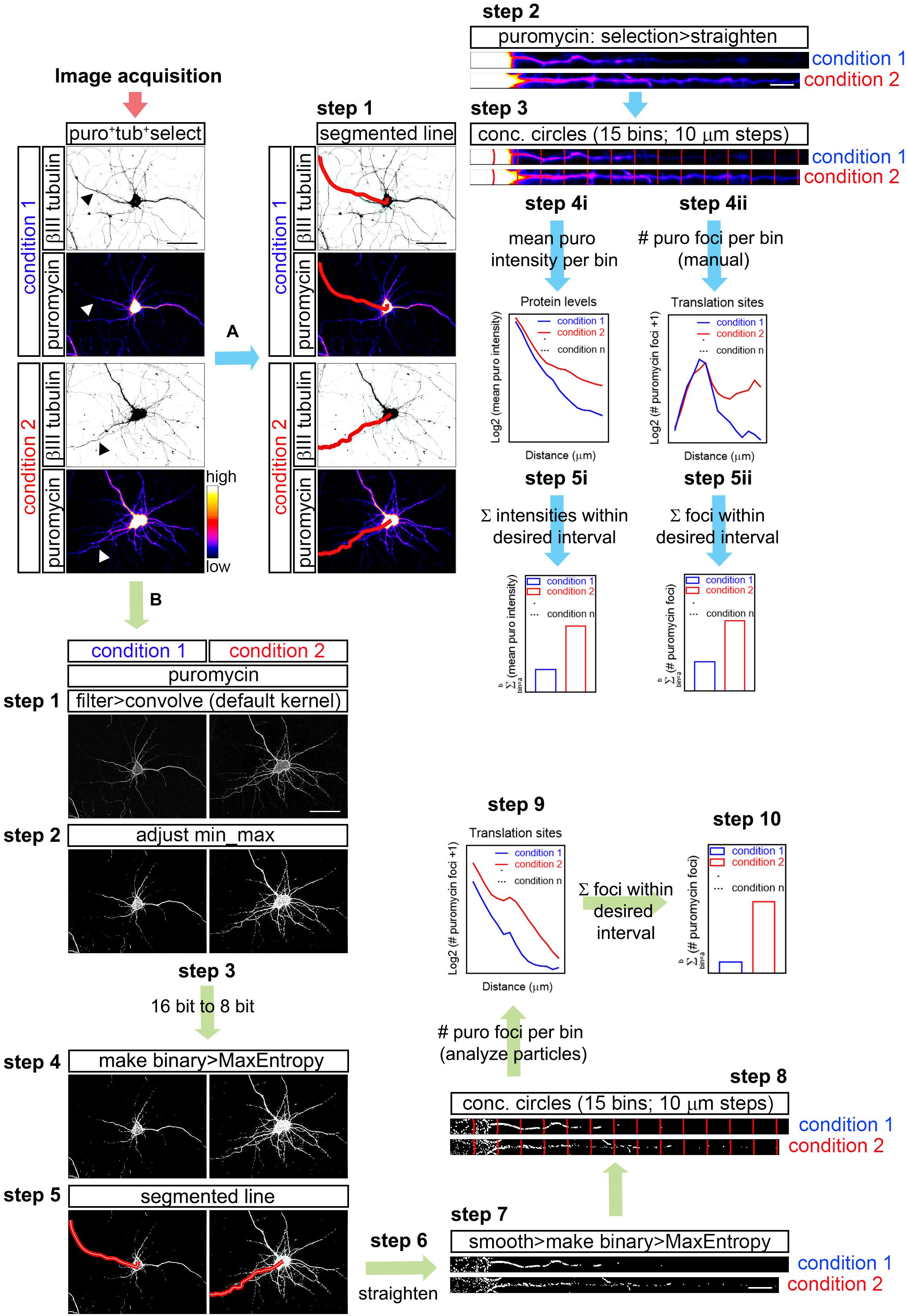
Frontiers Object Based Analyses In Fiji Imagej To Measure Local Rna Translation Sites In Neurites In Response To Ab1 42 Oligomers Neuroscience
![]()
Citing
Mij

Fiji Imagej With Batteries Included

Example Of Image Analyses Using Illastik And Imagej Fiji Illustrating Download Scientific Diagram

Fiji Imagej With Batteries Included
![]()
Citing
Image J Fiji Wolfson Bioimaging Facility University Of Bristol

Imagej Fiji Roi 1 Click Tools For Rapid Manual Image Annotations And Measurements